Membrane Master
Tamir Gonen
HHMI Janelia Farm
Published June 30, 2014
In college at the University of Auckland in New Zealand in the early 1990s, Tamir Gonen’s business classes bored him so much that he had his sister enroll him for his future classes. She put him on her track – medicine – and he never looked back.
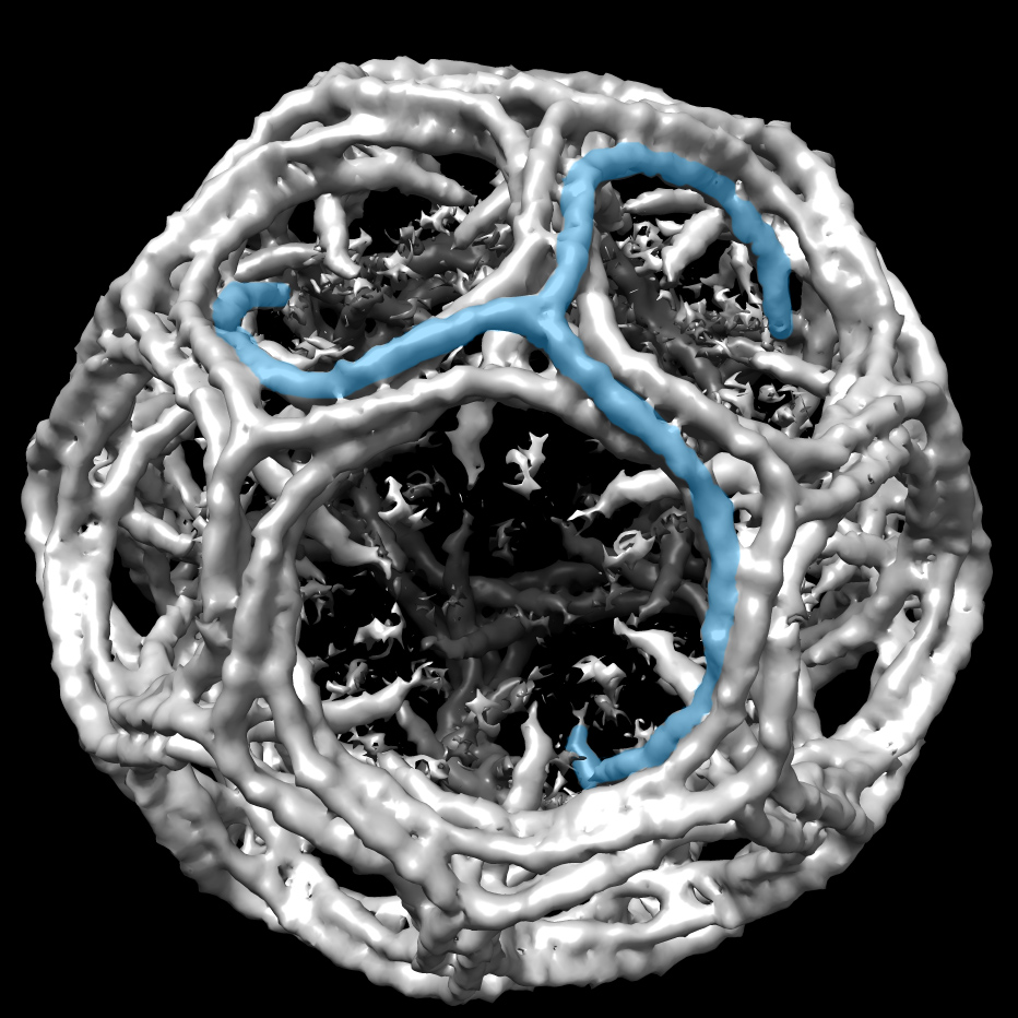
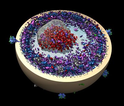
Gonen, now a Group Leader at the at Howard Hughes Medical Institute’s Janelia Research Campus, completed his bachelor’s degree in inorganic chemistry and biochemistry with First Class Honors. He focused his research on the lens of the eye, a clear tissue nourished not by blood vessels but by a network of membrane proteins that maintain homeostasis. Understanding these membrane proteins has been the driving force behind his work ever since.
His first project was to determine how cataracts develop, a question he only recently found a partial answer to. In 2012, he showed that interfering with phosphorylation of the membrane protein aquaporin-0 results in cataracts that are visually the same as those in a human patient.
After his Honors year, Gonen planned to move to Australia for his PhD. At the last minute, however, he decided to stay in Auckland to work with Ted Baker, a renowned crystallographer who had just relocated to the University of Auckland, and his current advisor Joerg Kistler, an expert in membrane proteins and electron microscopy.
With Baker, he started crystallizing membrane proteins and ended up with both MP20 and aquaporin-0 crystals. At the time, however, the local X-ray machine wasn’t working reliably, so he had a postdoc take his crystals with him on a trip to Stanford Synchrotron Radiation Lightsource. Inexplicably, the postdoc returned without diffraction data and without the crystals. “That was the day I left X-ray crystallography,” says Gonen.
Gonen completed his PhD by commuting — up to 5 times in one year — between Auckland and Boston, MA, where he worked with Tom Walz at Harvard Medical School to complete his studies of MP20 using electron microscopy.
To continue this work, he moved to the Walz lab as a post-doctoral fellow, where he completed the first atomic resolution structure of a mammalian membrane protein, aquaporin-0, using electron microscopy. “We could see water molecules and a belt of lipids around the protein, which was really transformative,” says Gonen, who also worked with Piotr Sliz to initiate the use of molecular replacement to phase electron diffraction data.
Gonen transitioned to the University of Washington, in Seattle, where he started his own lab focused on membrane proteins. In 2009 he joined HHMI as an Early Career Scientist and in 2011, HHMI offered him an opportunity to move to the Janelia Research Campus. He closed down his academic grants and moved to Virginia. “Time is probably the most important resource I have here,” he says. “It really allows us to go down exciting and risky avenues.”
Though Gonen has published widely on the structure and regulation of membrane proteins – using electron microscopy and X-ray crystallography – continued efforts are on pause for now in favor of developing new methods for studying them, such as MicroED. The method employs cryo-electron microscopy to diffract extremely small 3-D protein crystals, a million to 100 million times smaller in volume (100 times per side) than used in a traditional X-ray crystallography experiment.
He initially developed the method using soluble proteins and is now testing them on membrane proteins. “Many of the most important proteins we are interested in only form very small crystals,” says Gonen. “If you can use those small crystals to get meaningful data out of them, that makes some formerly impossible biology suddenly attainable.”
-- Elizabeth Dougherty